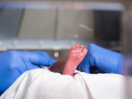
 Researchers have identified a gut-lung axis driven by intestinal antimicrobial peptide expression and mediated by the intestinal microbiota that is linked to lung injury in newborns.Lung development in the fetus occurs at low oxygen tension in the womb, but after a very premature birth, the partly developed lungs of the tiny infants experience far greater oxygen tensions even without the prolonged supplemental oxygen that is often required. This can produce well-known disastrous effects on the structure and function of the neonatal lung, causing the serious lung condition of bronchopulmonary dysplasia in high-risk premature infants.
Using a neonatal mouse model, researchers at the University of Alabama at Birmingham have shown that changes in the microbiota of the gut contribute to this dangerous lung injury. The changes occur because supraphysiologic oxygen exposure — 85 percent oxygen versus the normal 21 percent given to the newborn mice from the third to 14th days of life — suppresses expression of the normal host-derived antimicrobial peptides in the small intestine.
Antimicrobial peptides are known to play a key role in regulating intestinal microbiota through host defense against microbes. These small proteins produced by intestinal cells have a broad antimicrobial activity to kill or inhibit the growth of bacteria, fungi and viruses.
The novel finding of reduction of antimicrobial peptides upon exposure of the neonatal mice to supraphysiologic oxygen, in turn, led to altered composition of the intestinal microbiota in the neonatal mice, including increased abundance of bacteria in the genus Staphylococcus, as measured in the terminal ileum of the small intestine, the place where preterm infants are uniquely vulnerable to microbiome-mediated necrotizing enterocolitis.
Significantly, when the UAB researchers, led by Kent Willis, M.D., and Tamás Jilling, M.D., UAB Department of Pediatrics Division of Neonatology, gave supplemental lysozyme to the neonatal mice exposed to supraphysiologic oxygen, the augmented dietary lysozyme robustly altered the ileal microbiome to ameliorate the increase in Staphylococcus that had been seen after exposure to hyperoxia. Lysozyme is the most common antimicrobial peptide produced by the gut, and it is also highly expressed in human breast milk.
The functional consequence of this augmented dietary lysozyme was less lung injury, including improved lung structure and function after hyperoxia exposure in the neonatal mice. The dietary lysozyme also increased the gene expression of multiple antimicrobial peptides in the ileum through an apparent feed-forward mechanism, including a 2.4-fold increase in expression of the mouse lysozyme Lyz1 gene.
 Kent Willis, M.D.,
Photography: Lexi CoonTo test whether supraphysiologic oxygen directly suppresses antimicrobial peptide production by intestinal epithelial cells, collaborators in the Division of Newborn Medicine, Department of Pediatrics, Boston Children’s Hospital, Harvard Medical School, led by Amy O’Connell, M.D., Ph.D., grew intestinal organoids and exposed them to either hyperoxia (95 percent oxygen) or normal oxygen tension (21 percent) for 24 hours. Intestinal organoids are self-organized, three-dimensional structures derived from intestinal cells and grown in vitro to model the behavior of the original intestinal tissue.
After exposure to supraphysiologic oxygen, these intestinal organoids showed suppression of antimicrobial peptide genes, similar to changes seen in intestines of hyperoxia-exposed mice.
“In summary, we have described a gut-lung axis driven by intestinal antimicrobial peptide expression and mediated by the intestinal microbiota that influences hyperoxia-induced lung injury,” Willis said. “These murine and organoid experiments suggest that antimicrobial peptide expression represents a potential therapeutic target to modulate the intestinal microbiota and the response to lung injury. These results have implications for the clinical management of premature infants at high risk of developing bronchopulmonary dysplasia in neonatal care.”
Co-first authors of the study, “Antimicrobial peptides modulate lung injury by altering the intestinal microbiota,” published in the journal Microbiome, are Ahmed Abdelgawad and Teodora Nicola. Co-senior authors are Willis, Jilling and O’Connell.
Other co-authors are Isaac Martin, Brian A. Halloran, Kosuke Tanaka, Pankaj Jain, Changchun Ren, Charitharth V. Lal and Namasivayam Ambalavanan, UAB Department of Pediatrics Division of Neonatology; and Comfort Y. Adegboye, Harvard Medical School.
A video abstract of the study is available.
Support came from National Institutes of Health grants HL151907, HL156275, HL141652, DK120871, ES027697 and ES031559; the Harvard Digestive Disease Center NIH grant DK034854; the Kaul Pediatric Research Institute at Children’s of Alabama; and the Microbiome Center at UAB.
At UAB, Pediatrics is a department in the Marnix E. Heersink School of Medicine.