Case History
A 17-year-old young lady presenting with a pancreatic neck mass.
What is the most appropriate diagnosis?
- Nondiagnostic specimen
- Negative for malignant cells
- Atypical cells present
- Neoplastic cells present
Answer:
d. Neoplastic cells present


Brief explanation of the answer:
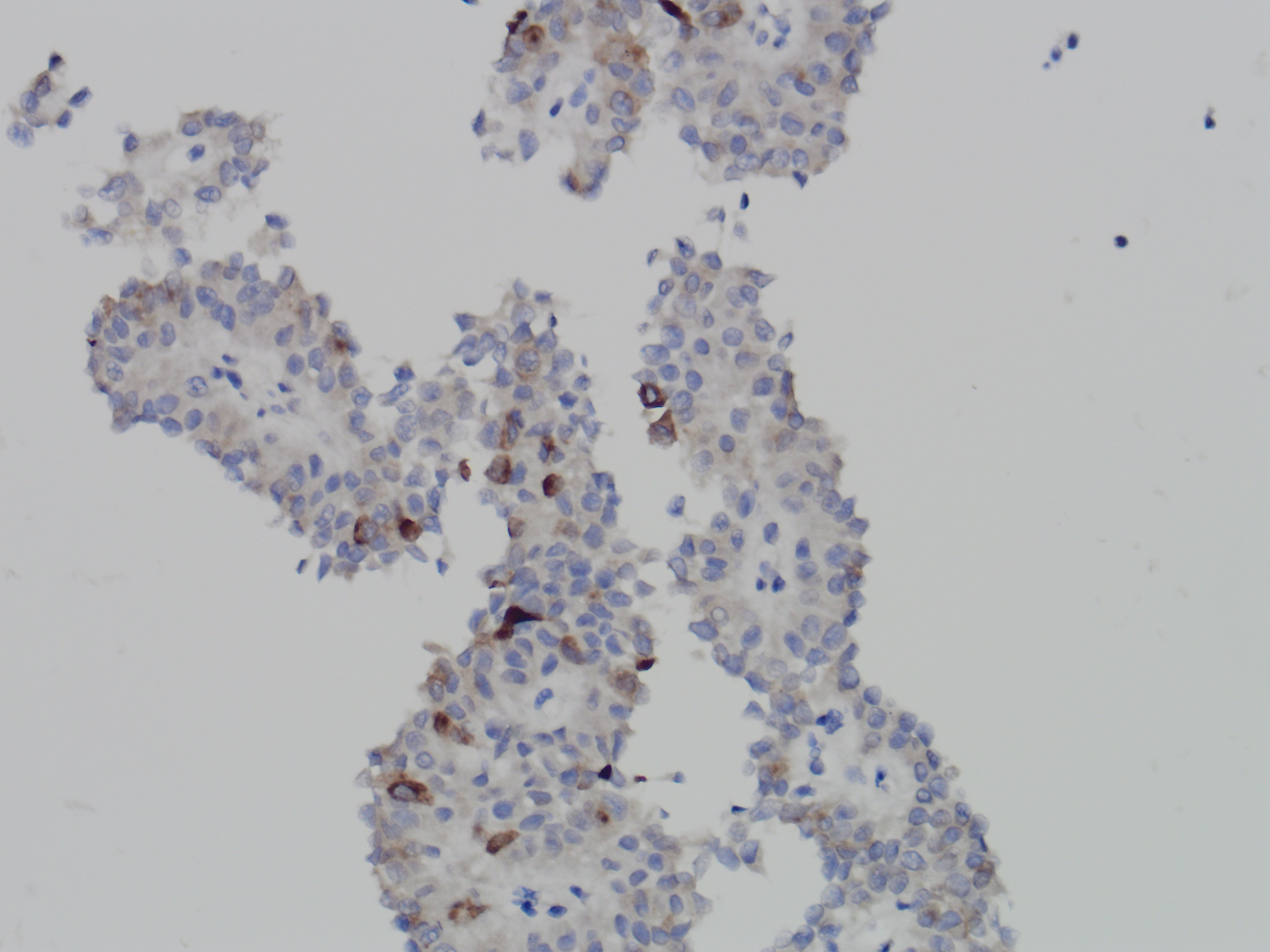
The specimen is richly cellular and consists of abundant monomorphic cuboidal nonmucinous cells, which are arranged as loosely cohesive groups and isolated cells (Figure 1). The neoplastic cells show delicate vacuolated cytoplasm with indistinct cell borders (Figure 2). These solid nests of loosely cohesive cells forming a cuff surrounding blood vessels result in a pseudopapillary architecture, which are well appreciated in the smear and cell block (Figure 3 and 4).
The differential diagnoses include pancreatic neuroendocrine tumor, solid pseudopapillary neoplasm and acinar cell carcinoma. By immunocytochemistry, the neoplastic cells show diffuse nuclear positivity for beta-catenin (Figure 5) and membranous positivity for CD10 (Figure 6). Also, the tumor cells are focally and weakly positive for synaptophysin (Figure 7). Taken together, the final diagnosis is solid-pseudopapillary neoplasm.
References:
Cytology, 4th Edition Diagnostic Principles and Clinical Correlates, by Edmund S. Cibas, M.D. and Barbara S. Ducatman, M.D.
Case contributed by: Yiqin Zuo, M.D., UAB Pathology.


