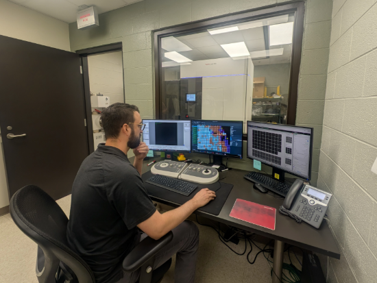
 Shane Rich-New, Ph.D., in the Cryo-EM Facility at UAB.In the basement of the Shelby Biomedical Research Building, one technology is helping UAB researchers understand the structures of proteins and other biological molecules and, in doing so, giving them insight into how to make unprecedented advancements in disease treatment.
“This kind of research is critical to truly understanding not only the basic science and the biology that we're interested in, but how the treatments that we design work and how we can make them better,” James Kizziah, Ph.D., associate director of the UAB Cryo-EM Facility.
Cryogenic electron microscopy, or cryo-EM for short is a powerful imaging technology that allows scientists to see the detailed shapes of proteins and other biological molecules by freezing them at extremely cold temperatures and imaging them with an electron microscope.
Freezing the samples preserves them in a near-natural state, letting researchers visualize how these molecules actually look and function inside living systems.
“Many diseases stem from proteins not working the way they’re supposed to—for example, enzymes that under- or overperform, or structures that fail to form or persist too long,” Kizziah said. “Proteins are fundamental to life, so when we understand their structures at the molecular and even atomic level, we can see not only how biology works, but how it goes wrong in disease. Cryo-EM lets us study those structures in extraordinary detail, which is essential not just for basic science, but for developing and improving treatments like drugs and antibodies by showing exactly how they interact with their targets.”
The UAB Cryo-EM facility is currently the only one of its kind in the state of Alabama, positioning UAB as a valuable resource advancing the study of disease and protein structures in unprecedented ways.
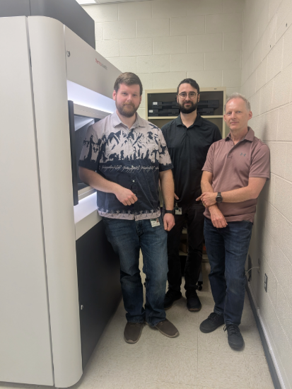 James Kizziah, Ph.D., Shane Rich-New, Ph.D., and Terje Dokland Ph.D., in the Cryo-EM Facility at UABForming a core facility
Cryo-EM at UAB leaped forward with the arrival of a new state-of-the-art microscope thanks to an NIH grant awarded in 2021 to Terje Dokland, Ph.D., professor in the Department of Microbiology and director of the Cryo-EM Facility. Prior to that, the lab was limited to an outdated microscope acquired back in 2006.
After Dokland was awarded the grant to acquire the new microscope, the idea of the lab becoming more of a shared resource emerged.
“We knew that we were upgrading and trying to be more of a core facility, serving many users,” Dokland said. “And so we knew we needed to have personnel."
To prepare for its installation, renovations took place to the lab in the Shelby Building, and Kizziah was hired to manage the facility before the new microscope was installed in August 2022. However, then came a setback.
A flood on Christmas Eve 2022 that affected many UAB facilities also flooded the basement of the Shelby Building, destroying three electron microscopes including the two used for cryo-EM in Dokland’s and Kizziah’s lab. Adjustments to the new microscope had been finalized just the day before. That put the lab out of commission for nine months, from January to September 2023, as replacements for the lost instruments were ordered.
“It's not the kind of instrument that you can just buy and pick up off a shelf,” Dokland said. “They have to actually build it for you.”
After the first replacement microscope was installed in September 2023, a silver lining came as the core facility was finally poised to take off.
“The silver lining for this is that in the interim period, the manufacturer had upgraded their systems,” Dokland said. “So, the replacement system was actually better than the original.”
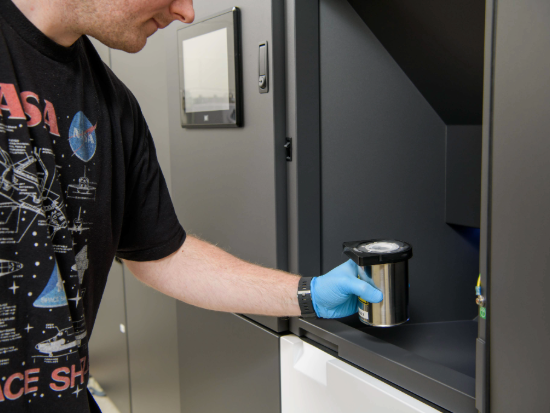 Giving a truer picture of protein structures—more efficiently
When considering the benefits of cryo-EM technology for analyzing protein structures, its efficiency compared to other methods is notable.
“It's progressed to a point where it can approach or match the structural detail provided by other techniques while avoiding some of the obstacles and limitations,” Kizziah said.
Compared to X-ray crystallography and nuclear magnetic resonance spectroscopy, cryo-EM can capture detailed images of proteins in more natural states in shorter time and with less material. This is significant because many important proteins are hard to produce in large amounts, and analyzing proteins in a more natural state gives scientists a better picture of how they function in the body.
“All these techniques still play an important role,” Kizziah said. “But recent advances in technology have made cryo-EM ideal for dealing with a variety of proteins and targets in a range of sizes and oftentimes within a time frame that may not be achievable by other techniques.”
Cryo-EM can produce approximately 10,000-15,000 images per day from up to 12 samples and can produce structures in approximately one to two months. The technique also works on a broader range of difficult targets, including large protein complexes, flexible or heterogeneous proteins, and membrane proteins, which are notoriously difficult to study.
These advantageous qualities have positioned cryo-EM for widespread use by researchers, as it is beneficial for multiple research areas that benefit from structural information.
In 2025, the Cryo-EM Facility hired Shane Rich-New, Ph.D., to serve as a manager and specialist in cryogenic electron tomography (cryo-ET), which focuses on understanding biological structures at the cellular scale, broadening the reach of cryo-EM at UAB.
“In the future, we aim to implement cryogenic focused ion-beam/scanning electron microscopy (cryo-FIB/SEM) for analysis of cell- and tissue-derived samples. Imagine being able to visualize a therapeutic agent or drug with an unknown mechanism in action in a cancer cell,” said Rich-New. “After treating your sample with the drug, you could use cryo-FIB/SEM to cut away a small window into what might be happening. It’s a field of research with incredible potential for UAB researchers.”
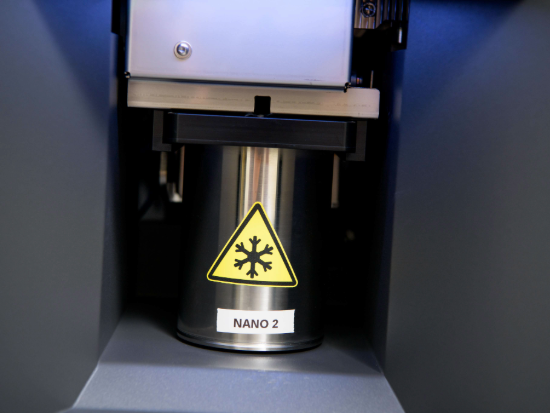 Speeding up scientific discovery
As a core facility, the cryo-EM lab is an available resource at UAB for various fields of study, from infectious diseases to neurological disorders to diabetes and beyond.
“About 80 percent of our users are in the School of Medicine,” Dokland said, adding, however, that the technology is available to anyone campus-wide.
Beyond UAB, the facility also receives requests to analyze samples sent from throughout the Southeast.
Since its implementation at UAB, the cryo-EM facility has already made strides in structural discovery, helping solve structures of the spike protein of SARS-CoV-2, immune system molecules, bacteriophage-related structures, and more.
As Kizziah and Dokland consider the future of the lab as a core facility at UAB, the goal has been to get the facility running as efficiently as possible while also training more researchers on how to use the technology.
“Anybody who is interested in applying cryo-EM to their project should reach out to us so we can work together on the grant writing process and help get their project started,” Kizziah said.