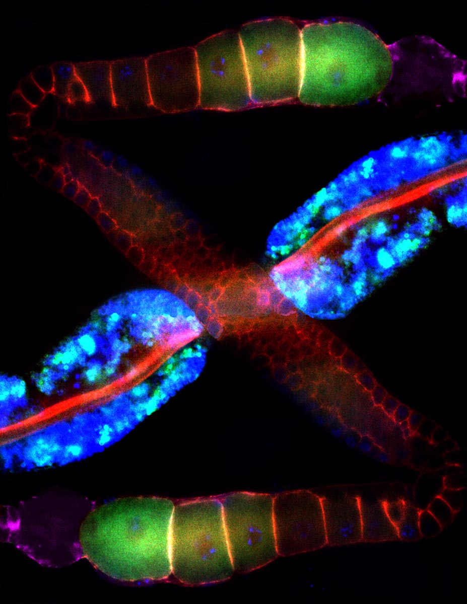
This image is a dissection of a species of worm called Caenorhabditis elegans (C. elegans), among the most famous of worms because it has made possible several discoveries in molecular biology. It did so by serving as a simple model of cellular processes conserved by evolution and still at work in humans.
The picture shows the worm's intestine running across the middle (in blue), which is working to provide fatty building blocks (e.g. omega-3 polyunsaturated fatty acid) to the worm's egg-producing cells (oocytes) in green. The egg-producing cells then convert the fatty acids into chemical cues called prostaglandins that help sperm find the sites (purple areas) where they can fertilize the eggs. C. elegans sperm in turn release a protein called major sperm protein (MSP), which tells the egg to prepare for fertilization.
Interestingly, the studies related to this image are providing key insights into how prostaglandins and MSP are made and function in humans. While MSP, for instance, was first found in worms and in connection with reproduction, it appears to have been put to work by human evolution in signaling roles in many cell types.
For instance, Michael Miller, Ph.D., associate professor in the UAB Department of Cell, Developmental and Integrative Biology, and creator of the attached image, last year published key work showing that MSP may be involved in the development of amyotrophic lateral sclerosis, or Lou Gehrig's disease. This unexpected connection may provide new approaches to diagnosing and treating the disease.
Note: if you have an amazing UAB research image you would like to share, please email me at gdw@uab.edu.