Four teams of University of Alabama at Birmingham researchers have been awarded National Science Foundation grants totaling $5.4 million meant to stimulate competitive research in regions of the country that are less able to compete for these research funds.
One research team supported by an NSF grant is in the College of Arts and Sciences’ Department of Chemistry, led by a polymer chemist who applies nanotechnology to biological and biomedical challenges. The three other grants will support basic neuroscience studies, one of UAB’s hallmark research strengths.
Lori McMahon, Ph.D., the Jarman F. Lowder Professor of Neuroscience, dean of the UAB Graduate School and director of the UAB Comprehensive Neuroscience Center, highlighted the three neuroscience EPSCoR grants at this fall’s Comprehensive Neuroscience Center retreat, calling them prestigious and competitive.
“UAB neuroscience has never had one NSF grant, and now we have three,” she said. “With the results of these grants, we can increase our funding beyond the National Institutes of Health.”
These four UAB grants, and one additional Alabama-related EPSCoR that supports research teams at the University of Alabama, Tuscaloosa, and the University of Mississippi, set a record, says Christopher Lawson, Ph.D., executive director of the Alabama EPSCoR program and a professor in the UAB Department of Physics.
“The state of Alabama now has more of the EPSCoR Track II grants than any other state; no state has ever had five.”
Only 25 states, two territories and one commonwealth — areas that receive much less NSF funding than the major research universities in the other 25 states — qualify to compete for the Experimental Program to Stimulate Competitive Research grants, known as EPSCoR. The EPSCoR Track II grants are meant to level the playing fields among have and have-not research states, and applicants must form collaborations with researchers from other EPSCoR states. This means the UAB teams have formed synergistic partnerships and collaborations across the Birmingham campus and with scientists in other states to foster regional research strength. 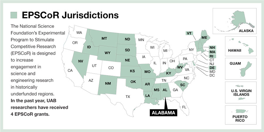
UAB neuroscience teams and their goals
The three neuroscience EPSCoR grants support a study to understand the initiation of epileptic brain seizures; a project to develop a new tool for optogenetics, which is the control of neural cells using light; and an effort to discover a universal rule for the relation between neural activity and increased blood flow in areas of the brain. In all three EPSCoR grants, UAB is a partner institution, and the lead institution is in another EPSCoR state.
“Several institutions reached out to UAB because we are so strong in neuroscience,” McMahon said. “There is a lot of UAB synergy around the three neuroscience EPSCoRs.”
McMahon says all three UAB neuroscience teams will meet regularly to share results and ideas.
Epileptic brain seizures
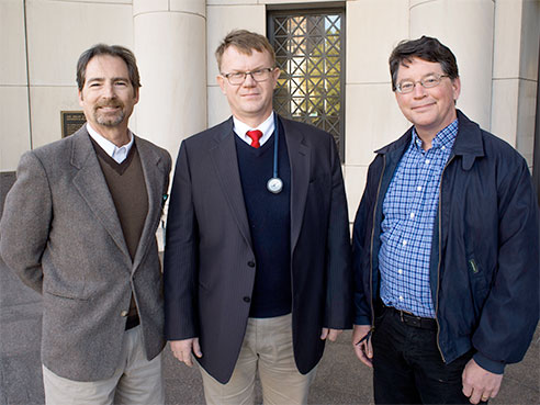 From left: Roy Martin, Jerzy P. Szaflarski and Timothy Gawne.The current approach for epilepsy surgery at UAB involves two surgeries. The first implants electrodes into a patient’s brain for a two-week period to map the location of the seizure onset zone in the brain. The second operation cuts out the onset zone.
“Our goal is to one day not need invasive monitoring,” said Sandipan Pati, M.D., assistant professor of neurology. “That would mean one surgery instead of two, and the patient would not have to stay in the hospital for two weeks.”
The UAB team in the epileptic brain seizure study — headed by Jerzy P. Szaflarski, M.D., Ph.D., professor of neurology — is developing and validating software for noninvasive brain mapping that will allow caregivers to locate that part of the brain that initiates seizures and locate those parts that function in memory.
“We will provide a road map for the surgeons — where to operate to remove the thumb-sized part of the brain that kicks off seizures, and what parts of the brain to avoid,” Pati said, “so that patients will have no added memory deficits after the operation.”
The investigators will use magnetoencephalography, or MEG, to map electrical activity using the magnetic fields produced by natural electrical currents produced by the brain. The magnetic forces are measured from the outside of the brain as the top of a patient’s head fits into the MEG device, which looks something like a beauty-shop hair drier on steroids. Pati says the UAB team has preliminary data about using the MEG to identify the onset zone by its hyper-excitation, without the need to wait for seizures.
Other UAB investigators in the study are Roy Martin, Ph.D., associate professor of neurology, and Timothy Gawne, Ph.D., associate professor of vision sciences. The lead institution for the study is Louisiana Tech University, and the University of Arkansas is also a partner in the study.
New tool for optogenetics
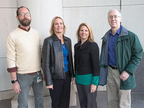 From left: Mark Bolding, Lynn Dobrunz, Lori McMahon and Gary Gray.Optogenetics uses light to control cells in living tissue, after light-sensitive ion channels are introduced into the cells by gene manipulation. Then light is sent into the brain on a fiber-optic cable inserted into the brain, to make neurons fire or to stop neurons from firing. This control helps researchers learn how the brain is wired and how it works.
The optogenetics project will create technology to control the light-sensitive ion channels using low-power X-rays, thus allowing control of neurons from outside the body.
McMahon is the UAB co-principal investigator in this project, which is led by Clemson University. Other partner institutions are the University of New Mexico and the University of South Carolina. The Clemson researchers will develop special nanoparticles that emit light in response to X-rays, the New Mexico researchers will genetically modify the light-sensitive ion channels so that they can bind the nanoparticles, and the UAB researchers will test how those nanoparticles disperse in the brain and how these particles, when activated by X-rays, can turn on and off brain circuits.
“We are about to do the first validation,” McMahon said. “The entire four years of the grant is developing the tool. Then we can use it in animal disease models for Alzheimer’s disease, Parkinson’s disease and anxiety. The goal is to understand brain circuitry using preclinical models of neurologic disease and models of neuropsychiatric illness.”
Other UAB investigators are Lynn Dobrunz, Ph.D., associate professor of neurobiology; Mark Bolding, Ph.D., assistant professor in the Division of Advanced Medical Imaging Research, Department of Radiology, and director of the Civitan International Neuroimaging Facility; Kazutoshi Nakazawa, M.D., Ph.D., associate professor of psychiatry and behavioral neurobiology; and Gary Gray, Ph.D., professor of chemistry.
Neural activity and blood flow
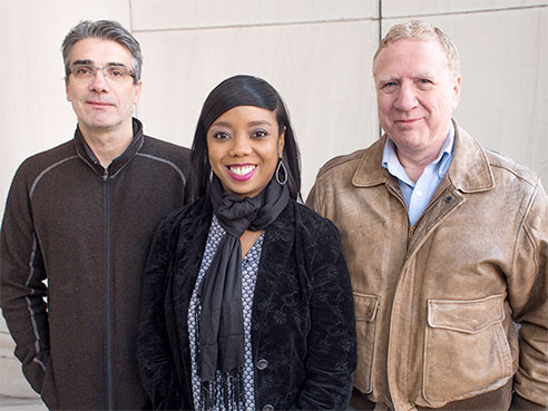 Jacques Wadiche, Farah Lubin and Paul Gamlin.It has long been known that neural activity is associated with increased blood flow, as the neurons need more oxygen and nutrients. In imaging with functional magnetic resonance, or fMRI, increased blood flow can be seen in parts of the brain as the subjects perform a task. But the resolution — both spatially and in time — is not sharp, and fMRI remains an indirect measure of neural activity.
The UAB team will use a very expensive infrared laser and microscope to peer beneath the surface of living brains to look at individual neurons and capillary beds.
“We will image cells in the brain using two-photon imaging,” said Paul Gamlin, Ph.D., professor of ophthalmology and UAB’s co-principal investigator in the neural activity and blood flow effort. “While monitoring neural activity, we will also monitor what the capillary bed is doing.”
“The question is, if we stimulate the cells, how precise is the blood flow change?”
Capillaries are the smallest blood vessels, bringing oxygen and nutrients to cells and taking away carbon dioxide and waste products. Networks of capillaries form tiny beds of vessels, and each capillary has a precapillary sphincter, a ring of muscle that can control blood flow, akin to crimping a garden hose to lessen water flow. It has been estimated that the human brain has 400 miles of capillaries, and that nearly every neuron in the brain has its own capillary.
Gamlin and others on the UAB team — Lawrence Sincich, Ph.D., assistant professor of vision sciences; Farah Lubin, Ph.D., associate professor of neurobiology; Jacques Wadiche, Ph.D., associate professor of neurobiology; and Yuhua Zhang, Ph.D., assistant professor of ophthalmology — can stimulate neurons nonphysiologically and see how the capillaries change. They will also look at physiological stimulation by shining light on one, two or three retinal cells in the eye and watching how neurons and capillaries in the visual cortex of the brain respond.
The lead institution in this project is the Medical University of South Carolina, and Furman University and the University of South Carolina, Beaufort, are partners in the study.
Detecting pollutants in Gulf Coast marine ecosystems
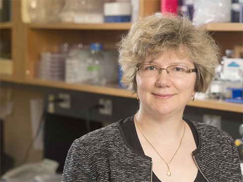 Eugenia KharlampievaThe Gulf Coast aquatic ecosystem hosts important fishing grounds and aquaculture, which co-exist with trading ports, off-shore oil wells and production industries. The fourth UAB EPSCoR is aimed at monitoring the water quality of this ecosystem, under the lead of the University of Southern Mississippi.
Ten researchers at six institutions in Alabama and Mississippi will develop advanced polymer-based, selective sensing technologies to detect and analyze pollutants.
The UAB investigator is Eugenia Kharlampieva, Ph.D., associate professor of polymer chemistry. She will design and synthesize three-dimensional porous hydrogel microparticles. These particles will be filled with sensing molecules created by Marco Bonizzoni, assistant professor of chemistry at the University of Alabama, Tuscaloosa. These hydrogels will eventually become devices that can sense polycyclic aromatic hydrocarbons in sea water.
“Sea water is a challenge because it is very rich in ions from all that salt,” Kharlampieva said. “It is hard to find the right technology that will work in that environment.”
Other researchers in the grant are developing sensitive and selective sensors to measure levels of carbon dioxide, nitrates and phosphates. The goal is new classes of seawater quality sensors that are faster, simpler and less costly. Other partner institutions in the grant are the University of Mississippi, Mississippi State University and Jackson State University.