Yuhua Zhang, Ph.D., was studying for his doctorate in precision metrology and instruments engineering at Tianjin University in China some 15 years ago when his focus quickly changed.
 |
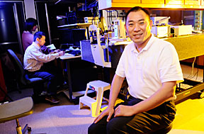
| UAB researchers, led by Assistant Professor of Ophthalmology Yuhua Zhang, have developed the fastest practical adaptive optics machine for the human eye. |
Zhang’s focus then shifted from learning to design cameras, telescopes and microscopes to ocular imaging, which he since has helped pioneer. Now, Zhang, an assistant professor of ophthalmology, has developed an adaptive-optics, 3-D imaging instrument that provides an unprecedented view of the human eye that ophthalmologists can use to identify and treat human-eye diseases.
“We have developed, to our knowledge, the fastest practical adaptive optics for the living human eye,” Zhang says of the adaptive-optics, scanning-laser ophthalmoscope (AOSLO), now in its third generation. “Imaging the retina in the living human eye with high resolution is important both for fundamental research and clinical patient care, and the development of this instrument and the excellent performance it has obtained has positioned UAB at the forefront of this emerging technology — technology that is available at only four other centers worldwide.”
The AOSLO will help ophthalmologists detect age-related macular degeneration, diabetic retinopathy and glaucoma, which will help them treat the diseases earlier and slow their progression. The instrument also provides unmatched images of rare eye diseases at the cellular level, which should give researchers and physicians a greater understanding of the pathology of these diseases.
“Dr. Zhang’s research truly is novel and cutting-edge,” says Lanning Kline, M.D., chair of the Department of Ophthalmology. “It provides a whole new way of looking both at normal anatomy and a variety of potentially blinding eye disorders.”
Zhang came to UAB in 2008 to establish an adaptive-optics program. Adaptive optics is a technology used to improve the performance of optical-imaging systems by reducing the effects of rapidly changing optical distortion. It first was created to help high-powered telescopes see clearly through the turbulent atmosphere in deep space.
The technology has been turned inward, and adaptive optics enables retinal imaging systems to compensate for aberrations on the cornea and lens of a patient. This leap in technology provides the ability to visualize living cells within the living human eye.
How it works
The UAB AOSLO is equipped with two state-of-the-art deformable mirrors that work together to correct the ocular aberration in virtually any human eye. By doing so, the AOSLO can image the retinal structure in the living human eye at the cellular level.
“The images are of unprecedented quality,” Zhang says, “enabling video images of microscopic blood flow and photoreceptors for diagnosis of eye diseases.”
Zhang says the AOSLO can obtain excellent retina images showing well-resolved cone photoreceptors in the macula, the central region of the retina that houses the densest pack of the cones, the cells forming the fine and color vision.
The ability to image retinal structure and function at the cellular level in such a high quality is a major achievement, Zhang says.
“With conventional clinical retinal imaging methods, we can not image these cells and the fine blood vessels,” Zhang says. “But with the AOSLO, we have the capability of directly imaging cones and the finest blood vessels. Furthermore, by controlling the intensity of the light beam forming the scanning pattern, the AOSLO can project clear visual stimuli with well-defined size, structure and intensity onto the retina. This will facilitate test of the retinal function. The high-resolution images will enable us to diagnose diseases at an earlier stage and provide sensitive evaluation of the treatment efficacy at the cellular level.”
AOSLO history
Zhang completed an earlier version of the AOSLO while working at the University of California at Berkeley with Austin Roorda, Ph.D., a professor of optometry. There they implanted a micro-electrical, mechanical system-based, adaptive optics into a scanning-laser ophthalmoscope and developed the AOSLO. Several problems limited the AOSLO’s use in clinic diagnosis; slow adaptive optics limited the image-acquisition speed and the ability of the machine to achieve its full image-quality potential.
Zhang’s goal when he first set up his UAB lab was to develop a new AOSLO and make it capable of high dynamic range, ocular-aberration compensation and high-speed imaging acquisition.
The AOSLO is ready for clinical studies now that goal has been achieved. Zhang is working with Cynthia Owsley, Ph.D., Christopher Girkin, M.D., Christine Curcio, Ph.D., and Douglas Witherspoon, M.D., to study age-related macular degeneration and primary open-angle glaucoma.
“They all are world-renown scientists and physicians,” Zhang says. “We also will study other common medical and neurologic conditions, including hypertension, diabetes, multiple sclerosis and Alzheimer’s disease.”
Zhang says the support from Kline and career-development mentors and collaborators Owsley, Girkin, Curcio and Judith Kapp, Ph.D., has made the development of the AOSLO possible. Also crucial to the effort, he says, was the support from Vice President of Research and Economic Development Richard Marchase, Ph.D., and Assistant Vice President of Research Charles Prince, Ph.D., and Vision Science Research Center Director Kent Keyser, Ph.D.
“The support I’ve been given has been unbelievable,” he says.
Zhang’s work is supported by grants from the EyeSight Foundation of Alabama, International Retina Research Foundation, Buck Trust of Alabama and Songs for Sight.